Utah's first FreeMax MRI Increases Research and Clinical Imaging Capabilities
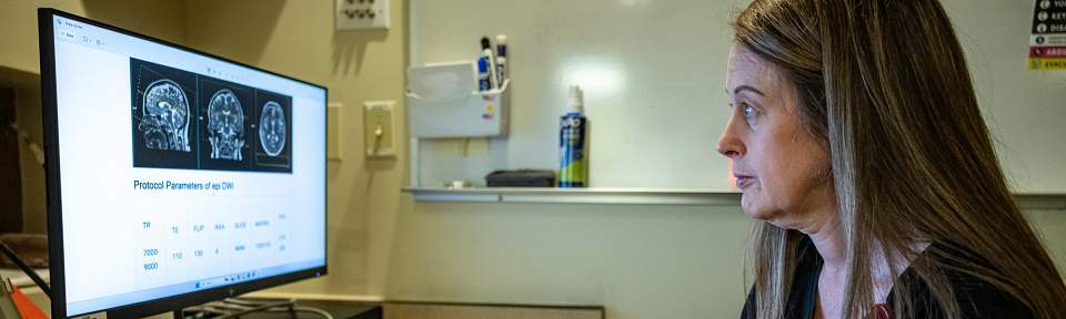
The Utah MRI Research Center (UMRC) Department of Radiology and Imaging Sciences has completed the installation of Utah's first Siemens Free.Max—an MRI scanner that operates at a field strength of just 0.55 Tesla, a significant shift from the standard 1.5T or 3T clinical MRI units. Despite its lower magnetic field, the Free.Max delivers high-quality diagnostic images, thanks to its integration of state-of-the-art artificial intelligence that enhances image sharpness, clarity, and overall quality. The scanner also supports green energy solutions for the University by reducing the use of coolant and helium from over 1,000 liters to 0.7 liters.
UMRC began exploring the potential of low-field MRI technology several years ago, recognizing its promise as a cost-effective and versatile solution to meet both clinical and research needs.
Clinically, the scanner is versatile, but also allows imaging for patient populations that were previously limited or excluded from MRI imaging. Patients with metallic joint replacements, or orthopedic hardware—historically difficult to image—can now receive detailed MRI scans to support accurate diagnoses. Another patient-focused advantage is the scanner's 80 cm bore, the largest at the University. This wider design increases comfort for patients who experience claustrophobia or discomfort in traditional MRI environments, leading to less anxiety and failed scans.

"The installation of Utah's first AI-enhanced low-field MRI represents a transformative moment for our clinical practice and research mission," said Sam Finlayson, MD, MPH, MBA, Chief Clinical Officer of University of Utah Health. “This new equipment expands access to essential diagnostic imaging, advances our research capabilities, and provides a new tool for training the next generation of physicians and researchers on leading-edge, sustainable technology."
The MAGETOM Free.Max has begun taking clinical scans and is scheduled to be fully operational by July 1, 2025. Currently, the scanner has been tested for shoulders, knees, ankles, routine brain images, spines, and orthopedic hardware. UMRC is currently testing imaging with contrast and other additional limited scans. UMRC anticipates a high volume of scans focused on patients with hardware, leveraging the system's unique strengths.
The decision to adopt the Free.Max was driven not only by its clinical potential, but also by the opportunity it presents for innovative research collaborations. Several research groups are preparing to transition their work to this new platform.
In partnership with the Coil Lab, Rock Hadley, PhD, is initiating projects focused on developing next-generation MRI coil technologies for the Free.Max. Additionally, teams are exploring the integration of low-field MRI with ultrasound applications, further expanding the scanner's utility across modalities.
“The MRI research team at the University of Utah is excited to leverage the advanced features of the Siemens Free.Max—particularly its larger bore size and lower field strength—to push the boundaries of image-guided procedures, improve cardiovascular health, and, most importantly, enable patient-centered imaging needs including broadening the patient population that can benefit from MRI," said Allison Payne, PhD, Vice Chair for Research in the Department of Radiology and Imaging Sciences.
“The installation of the Siemens Free.Max marks a major milestone for the university and the state of Utah by positioning UMRC at the forefront of low-field MRII innovation." said Satoshi Minoshima, MD, PhD, the Chair of the Department. With its advanced AI capabilities, patient-centric design, and research potential, this technology will significantly enhance both clinical offerings and research capabilities.