CQCI Pre-Clinical Research Imaging - Resources
The CQCI preclinical research facilities include a basic science lab, cold chemistry and radiochemistry labs, and small animal imaging lab within the HCI vivarium. These facilities are used for developing novel diagnostic imaging compounds and cancer therapeutics utilizing a wide range of cancer rodent models. CQCI is also the North American Mediso Preclinical Imaging Center of Excellence and has the first installed NanoScan 3T PET/MRI in the United States.
The major instrumentation in these labs are listed below:
-
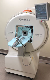
Mediso nanoScan SPECT/CT
-
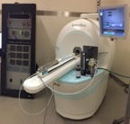
Mediso nanoScan 1T PET/MRI
-
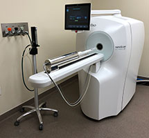
Mediso nanoScan 3T PET/MRI
-
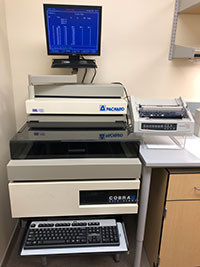
Packard Cobra II Auto Gamma Counter
-
HM 560 CryoStar
-
BAS-5000 Image Analysis System
Mediso NanoScan SPECT/CT
The nanoScan SPECT/CT system is a high-speed, high resolution device for both focused and whole-body imaging of small animals. The integrated, ultra-fast, low-dose and variable FOV micro-CT provides automatically registered anatomical information for fusion and corrections. The preclinical SPECT/CT system supports quantitative dynamic imaging with full list mode acquisition format. Gated possibilities are offered for both modalities.
SPECT Features:
- Up to 250 mm transaxial FOV
- 300 μm spatial resolution
- 13 000 cps/MBq sensitivity
- Up to 144 pinholes
X-Ray CT Features:
- 80 W/1 mA X-ray tube power
- <10 mGy exposure CT dose
- 2-12 cm variable TFOV
- x 7,6 zoom
- <10 μm isotropic voxel size
Mediso NanoScan 1T PET/MRI
MRI Features:
- 1 Tesla Permanent magnet
- Gradient strength of 450 mT/m
- <= 100 mm spatial resolution
- Integrated gradient coil
- Integrated RF and magnetic shielding
PET Features:
- LYSO crystal full ring geometry
- Transaxial FOV: up to 12 cm
- Aperture: up to 16 cm
- Spatial resolution: up to 0.3 mm3
Mediso NanoScan 3T PET/MRI
MRI Features:
- 3 Tesla cryogen-free magnet
- Gradient strength > 450 mT/m
- +/- 0.5 ppm over 50 mm DSV
- Stability: <0.05 ppm/hour
- <= 50 mm spatial resolution
- Integrated gradient coil
- Integrated RF and magnetic shielding
PET Features:
- LYSO crystal full ring geometry
- Transaxial FOV: up to 12 cm
- Aperture: up to 16 cm
- Spatial resolution: up to 0.3 mm3
Packard Cobra II Auto Gamma Counter
This automatic, multidetector gamma counter is designed for detecting and measuring gamma radiation. With a ten-detector system for high throughput screening, the Cobra II has the tube capacity for 750 to 1000, depending on tube size. The Cobra II is utilized for counting large batches of ex vivo tissue samples using short-lived radioisotopes.
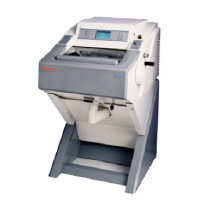
HM 560 CryoStar
The basic sciences laboratory has a Microm HM560 CryoStar for specimen preparation. This cryomicrotome is an electronic, motorized cryostat with retraction that incorporates a unique refrigeration system for independent specimen and knife cooling.
Other features include the following:
- Separate knife temperature control to -35°C
- Object temperature separately controlled to -50°C
- Integrated Peltier to -60°C
- Section thickness from 0.5 to 100µm
- Automatic Cryo Approach (ACA) system for exact and safe approach of specimen towards the knife
- Automatic cutting window
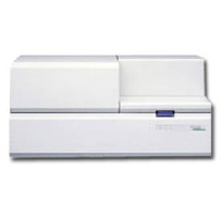
BAS-5000 Image Analysis System
The Center for Quantitative Cancer Imaging has as part of its basic science imaging infrastructure a Fuji BAS-5000 Phosphor imager. This device offers extremely high resolution (25 μm) and image quality. It uses Fujifilm’s unique confocal laser and light-collecting optics. The BAS-5000 is amenable to fine-structure studies of a wide variety of tissue samples. With a dynamic range up to five orders of magnitude and a pixel size as small as 25 μm, the system allows for very high-resolution quantitative autoradiography studies. The system can provide rapid scan times. A 20 x 25 cm imaging plate can be scanned at 50 μm in as little as five minutes. The system includes software for region of interest (ROI) analysis and quantitative assessment of tracer accumulation. The system can image all PET isotopes as well as C-14 and tritium. The system also has the capabilities to perform various optical-based assays, including 2D electrophoresis and thin layer chromatography.
Contact Us
Jeffrey Yap, PhD
jeffrey.yap@hci.utah.edu
801-213-5650
