CQCI Clinical Research Imaging - Resources
Molecular Imaging PET/CT Suite
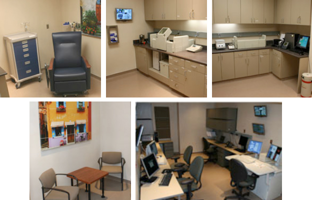
A dedicated molecular imaging PET/CT research suite is located adjacent to the cyclotron and radiochemistry laboratory. This suite is 2,300 square feet of dedicated research space for imaging both human research subjects and large animal imaging when needed.
This space has a large room with the GE Discovery ST PET/CT system (8 slice CT) and another large room with the GE 710 PET/CT scanner (64 slice CT). There is a large common control room for the operation of the two scanners. There is an additional work area for image processing. In addition there is an electronics room, patient waiting area, patient uptake, patient restroom, and hot lab. The hot lab contains shielded storage areas for radioactive materials and radioactive waste. There is also a Capintec dose calibrator, radiation safety monitoring equipment, Capintec single sample well counter, and a PerkinElmer Wizard2 Gamma Counter. Additional equipment includes a centrifuge and a glucometer. The image process equipment consists of three GE Unix workstations, a GE AW workstation, iMac computer, PCs, and a high-performance server (Universal 2U Dual Quad-core Xeon with 4 terabytes of disk). In addition, there is a high-end workstation for processing preclinical images.
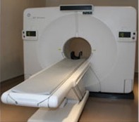
GE Discovery ST
The GE Discovery ST is an advanced PET/CT scanner that combines PET and CT into one inherently registered image. The system includes the Discovery Dimension operator’s environment. The fused images capture essential information from each imaging technology to shed new light on oncology, cardiology, and neurology. The scanner is the only currently available tomograph optimized for both 3D and 2D imaging with full septa for optimal imaging of patients of all sizes. Isotropic crystal design also accurately shows lesion size and shape. The device features 30 mm thick BGO detector crystals, selected for effectiveness with cancer radiotracers for the highest stopping power. It also contains square quad photomultiplier tubes specially engineered to boost the detector’s efficiency at collecting image information significantly. It also includes a full-featured eight-slice GE LightSpeed multi-slice CT with attenuation correction. The Discovery ST can also be used as a stand-alone CT. It simultaneously acquires multiple adjacent slices of anatomy with solid-state GE HiLight Matrix detectors that include high X-ray stopping power, low afterglow, and z-axis uniformity and transparency. The tomograph has optimized image quality that covers a full range of applications with speeds of up to 0.5 seconds and variable scan speeds adjustable in 0.1-second increments. It features a 100 cm bore length and a large 70 cm bore width, minimizing patient claustrophobia. Connectivity to various systems is through PET and CT DICOM 3.0. The system includes both respiratory and cardiac gating capabilities. It also includes two GE Advantage Workstations (AWS) for image review and processing.
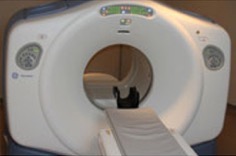
GE Discover710 PET/CT
The Discovery710 PET/CT scanner was installed in early 2013.The tomography is the latest high-performance scanner available from General Electric Healthcare. The scanner employs 13,824 LYSO crystals in 24 detector rings to provide 47 slices per bed position. The spatial resolution is ~5mm FWHM. The scanner operates in a time of flight (ToF) imaging mode and has a variety of reconstruction options, including SharpIR technology that utilizes point spread function correction to improve lesion detection with high resolution. The operating modes include respiratory gating of both CT and PET images and total body scans are possible for patient heights over six feet in a single scan. The CT for this system is a 64-slice BrightSpeed Elite system with fast helical volume imaging with low-dose capability and ASiR CT adaptive reconstruction algorithms for high-quality, low-noise images at low mA. Reconstruction matrix size is 512X512 with 1024X1024 display capability. Workstations for this PET/CT system include complete packages of software for display and analysis software for oncologic and neurology applications.
Image/Data Analysis Lab
The image process equipment consists of three GE Unix workstations, two GE AW workstations, iMac computer, PCs, and a high-performance server (Universal 2U Dual Quad-core Xeon with 4 terabytes of disk). The CQCI also uses the MIM Encore image viewing and analysis software. In addition, there is a high-end workstation for processing preclinical images.
Research Image Archive
The CQCI has a dedicated research PACS system for archiving and accessing research images. The NUMAstore PACS system is configured through an HP six-core Dual Xenon CPU server and has a 6TB storage array of 300GB SAS HDD disks that are configured in RAID 6 with hot swap drives and redundant power supplies. The hardware is expandable to 18TB. The operating system is Microsoft Windows 2008 Server OS. The system has a high capacity 2 Dual Port NIC, which connects via a secured network to scanners and workstations within the molecular imaging department. There is also a pathway to securely import images from the University enterprise-wide PACS system. Data is stored with loss-less compression and with incremental backup on high-density 800GB SAS tapes on a weekly basis. There is full system backup monthly. The hardware is housed in a main server room at Huntsman Cancer Institute (HCI) with security, backup power, and a non-water fire suppression system. The system interfaces with two PET scanners, a PMOD workstation, and six imaging workstations.
Utah Center for Advanced Imaging Research (UCAIR)
The Utah Center for Advanced Imaging Research (UCAIR) aims to advance medical imaging technology by applying the tools of physics, mathematics, and engineering. UCAIR is primarily housed at the Imaging and Neurosciences Center. Dr. Hoffman is the medical director of UCAIR. This aim is achieved by providing the following:
- Infrastructure and support for grant submittal and management to foster all aspects of research on medical imaging technology
- Imaging expertise to all departments within the university campus. This includes instrumentation as well as Physics and Radiology support for University projects in areas of high-end human imaging.
The Imaging and Neurosciences Center building currently houses UCAIR.
Contact Us
Jeffrey Yap, PhD
jeffrey.yap@hci.utah.edu
801-213-5650


